Gas Chromatography Mass Spectrometry GC:MS.
─
John Holmes Mass Spectrometry Facility
Hydrosol Creation and Analysis
124 D’iorio Hall
University of Ottawa
Overview
This document will instruct you in the basic principles of GC:MS and the operating procedures.
Goals
- To understand how the GC:MS works, so you can create a method to run it.
- To run a sample and obtain good separation of the chemical components on the column.
- To use the Data Analysis software to identify chemical components and evaluate the precision and accuracy.
Specifications
The instruments available are Agilent GC:MS’s there are 3 of them.
As stated in the name there are two parts to the instrument.
The Gas Chromatography section, we have models HP 6890, 7820A and 6890N Agilent, Hewlett Packard was bought by Agilent.
The Mass Spectrometers are all Electron Impact Sources (EI) with a single Quadrupole (Quad) mass filter. Models: 5975 MSD and 5975C.
GC/MS Instruments, GC/MS Systems, GC/MS Analysis | Agilent
Brief Summary of Procedure
A method is created in the GC software and uploaded to the instrument.
A 1 ul dilute fully soluble solution of hydrosol created as previously stated and refined using solid phase extraction cartridge, is injected into the inlet of the GC section of the instrument. The method is started and the GC oven ramp heats the column that contains the hydrosol components that were vaporized in the inlet and carried to the column by helium carrier gas. As the oven gets hotter the individual chemical components will elute from the GC column according to their boiling points and interactions with the column. The molecules enter the ion source of the mass spectrometer where they are ionized and fragment. The molecular ion (M+) and fragments are then separated by the quadrupole mass spectrometer and arrive at the detector where their mass to charge is graphed against their intensity, this is called the mass spectrum. Each molecule will have a different mass spectra, thus this graph becomes the individual fingerprint of that molecule and can be compared to a standard mass spectrum of the pure compound that is contained in a database within the software of the instrument, data from thousands of molecules are contained in the database. Electron Impact Mass Spectra are also contained in the Nist Chemistry Webbook, NIST Chemistry WebBook..
Milestones
- Create Method in the GC
A number of parameters can be controlled in the gas chromatography section. These include:
Flow rate of the helium carrier gas.
Inlet temperature needs to be hot enough to vaporize all your molecules but not too hot to decompose them.
Temperature ramp on the GC column needs to start at the first appearance of a compound, needs to not start during the solvent elution and needs to finish just after the last compound and go slow enough to separate out all your components and not too slow to last hours.
- Create Method in the MS
The MS method can be altered to allow you to just select particular ions and monitor just these, this is called a single ion monitoring scan, this is employed for very specific methods generally the mass spec is set to scan from 18-614 da. Sometimes we will start the mass at 33da so as not to record air. We also set the mass spectrometer to off for the first 3-4 minutes if the solvent will appear at that time. Solvent is 999 times more concentrated than our sample and it has a low boiling point so will pass through the entire apparatus in the first few minutes. We do not want or need to record it.
Once our method is created we can name it and save it to the computer and make sure it's loaded into the GC and then we are ready to run.
- Analyze the data.
Become familiar with the gas chromatogram and how to access the mass spectrum under each chromatogram peak and use the database to identify the compound.
Details
- Create Method in the GC
1. What is the boiling point of the compounds you are interested in?
What is the highest boiling point of the compounds in your sample?
What is the highest temperature you can run your column to?
For instance a non-polar HP 5 column is 325°C Column Types (I would not run above 300°C on a regular basis) but a polar column is only 250°C. If you run above these temperatures the column will start to deteriorate, which means column material will be carried to the mass spec and will either (or both) show up as a high base line with mass spec peaks consistent with the column material or there will be chromatogram peaks that show the mass spec of the column material. Know what the mass spec of column bleed looks like so you can identify it. Column and Septum Bleed
The column is inside the GC oven and looks like this:

The inlet is seen at the top left and the column emerges from there, it is 30 m and is wound around just in front of the heater fan, it enters the mass spectrometer on the left wall. It is often hot, be careful.
Run the oven temperature so that all the material is carried through the column in the run, even compounds you are not interested in. If you do not, material will stay on the column and contaminate your next run. Running a blank is good practice to make sure everything is clean.
The settings for the oven temperature can be accessed in the software under the oven icon that looks like a column.
When you click on the oven icon another page opens where you can set the oven temperature ramp.

Most of the GC settings can be accessed from here.
From left to right the icons mean:
First is the ALS, this chooses the injection method, which could be manual or autosampler.
There aren’t any valves on our instruments but there could be gas valves.
Next is the inlet, we will deal with that next.
AUX Heaters can be other components that need heat, could be a gas valve or the line from the GC to the MS.
2. The inlet needs to be at a temperature that vaporizes all the compounds, what is that?
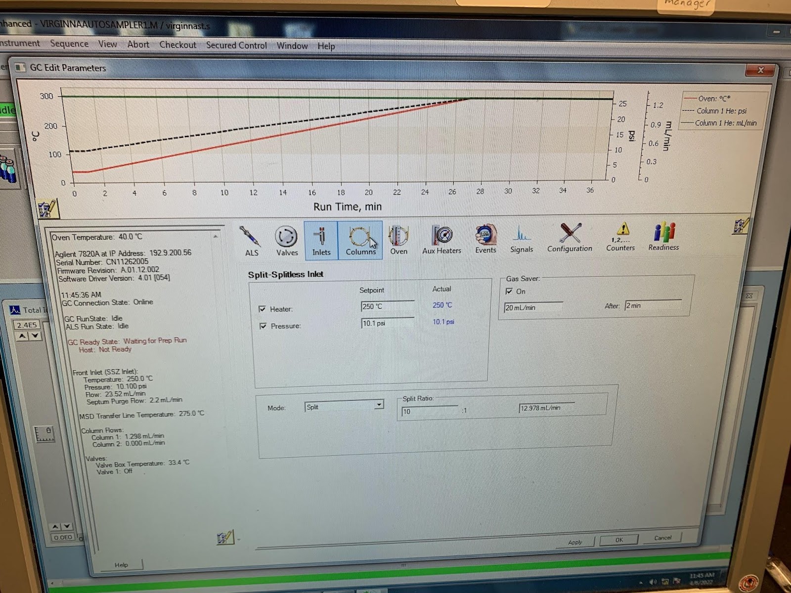
The inlet settings in the program that operates and runs the GC:MS can be located by clicking with the mouse on the oven icon in the software, followed by the inlet icon. inlet

The temperature of the inlet in this method is set to 200°C, the samples this method is designed to analyze are not very volatile organic compounds, unlike the terpenes of essential oils and hydrosols.. The temperature in the inlet needs to vapourize all your compounds without decomposing them.
3. The inlet split is set here. The sample is not all sent to the column, some is sent to waste and only some to the column.
What split ratio shall I use?
A large split is chosen for concentrated samples, GC:MS is a sensitive analysis and concentrations exceeding 1mg/ml should be diluted.
4. The helium carrier gas pressure is also seen here, this is a good pressure for separation. Helium gas can be run at constant flow or constant pressure, there are advantages and disadvantages for each. This is set in the column icon.
Knowing or making an educated guestimate of the boiling points of your compounds will help in deciding all these quantities but trial and error is often needed to design the best method. The pressure that the column is under will affect the boiling points of your compounds.
What pressure/ flow rate and temperature gradient shall I run the helium carrier gas at?
Again, 10psi and 12°C per minute are good starting points. Most people run a constant flow method which will determine the helium pressure. 1-2 ml/min is a good suggestion. This you can alter to get the best separation of the chromatogram peaks and the faster run, balance. Flow rates are controlled in the column settings.
Note: For non-polar columns boiling point is the most important criteria for the order of emergence from the column, however for a more polar column the interaction of the column with the molecular structure will play a very significant role in peak order.
- Create Method in the GC, example:
0.2 µL of essential oil was injected into the inlet of an Agilent 7820A Gas chromatograph. The inlet was heated to 150°C to volatilize the oil before being carried to the column by the helium gas. The carrier gas was at a pressure of 10.1 psi and a flow rate of 16.5 ml/min. The inlet split ratio was set to 10:1 meaning approximately 9% of the volatilized sample entered the column and the rest was sent to waste. This is done to avoid overloading the column as well as saturating the detector of the MS instrument. A non-polar, 30m long HP5-MS chromatography column with a 250 µm diameter and 0.25 µm pore size from Agilent (Mississauga, Ontario, Canada) was used to separate the compounds. The stationary phase of the column is 95% methylated siloxane with 5% phenyl siloxane allows better separation of non-polar analytes such as the terpenoids found in essential oils. The oven temperature was held at 40°C for one minute then increased to 300°C over the next thirteen minutes at a rate of 20°C/min and then held constant at 300°C for 2 minutes. This low starting temperature was used since essential oils are very volatile and most compounds elute at low temperatures. The samples elute primarily in order of their relative volatilities but the Van Der Waals interactions between the compounds and the non-polar stationary phase improves the separation.
- Create Method in the MS
This is often not tampered with but it does contain the timing so the solvent front is not seen.

This is accessed via the icon that looks like a quadrupole next to the column icon. The solvent delay in the first box. Nothing else tends to get altered here.
Voltages on the quadrupole are optimized during a tune and the MS has a tuning solution PFTBA situated near the source which it uses. Perfluorotributylamine - Wikipedia
The source is an electron impact source which is set to the standardized 70eV that everyone uses. The mass filter is a single quadruple. Quad
The GC and MS settings can also be accessed in the software using the tabs, see below.

- Analyze the data.
The data is viewed and manipulated in a different program than the one that operates the instrument. The program is called “Enhanced Data Analysis”.

You load the sample run using the file tab.
You will need the retention times on each of the chromatogram peaks, seen in the top window and numerically in the bottom window. The bottom window is called the “percentage report” and shows the retentions times and area beneath each peak and the percentage of each peak relative to all the peaks This is obtained by clicking on the chromatogram tab and selecting PERCENTAGE REPORT.
To acquire the mass spectrum and identify each compound represented by each peak it is necessary to zoom in on the individual peaks and sample the area within a peak using the mouse.

When you do this the bottom window will show the mass spectrum for that peak.
If you double click on the mass spectrum it will search the database for a matching spectra.
When it finds a few it will produce a window showing you its choices.

The box tells you what database it used and what compounds it matched your mass spectrum too. It tells you how well they match 100% being perfect and it shows you the database spectra too.
This can also be done automatically under the spectra tab, choose Library Search Report, there are two options summary and detailed. The software will automatically search every peak and match the mass spectrum and produce a PDF that can be saved or printed.
The skill is in evaluating the software choices.
 Chromatogram.

Percentage Report

Mass Spectrum of Chromatogram peak at 8.629 mins is matched 98% by the database to Citronellal. This essential oil is citronella, so we can be sure that that is what it is.


